| Info
Sheets |
| | | | | | | | | | | | | | | | | | | | | | | | |
 | Out-
side |
| | | | |
|
| | | | |
Result : Searchterm 'mra' found in 2 terms [ ] and 57 definitions [ ] and 57 definitions [ ] ]
| | previous 36 - 40 (of 59) nextResult Pages : [1] [2 3 4 5 6 7 8 9 10 11 12] |  | |  | Searchterm 'mra' was also found in the following services: | | | | |
| |  |
| |
|
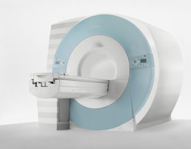
From Philips Medical Systems;
the Intera-family offers with this member a wide range of possibilities, efficiency and a ergonomic and intuitive serving-platform. Also available as Intera CV for cardiac and Intera I/T for interventional MR procedures.
The scanners are also equipped with SENSE technology, which is essential for high-quality contrast enhanced magnetic resonance angiography, interactive cardiac MR and diffusion tensor imaging ( DTI) fiber tracking.
The increased accuracy and clarity of MR scans obtained with this technology allow for faster and more accurate diagnosis of potential problems like patient friendliness and expands the breadth of applications including cardiology, oncology and interventional MR.
Device Information and Specification
CLINICAL APPLICATION
Whole body
CONFIGURATION
Short bore compact
Standard: head, body, C1, C3; Optional: Small joint, flex-E, flex-R, endocavitary (L and S), dual TMJ, knee, neck, T/L spine, breast; Optional phased array: Spine, pediatric, 3rd party connector; Optional SENSE coils: Flex-S-M-L, flex body, flex cardiac
SE, Modified-SE ( TSE), IR (T1, T2, PD), STIR, FLAIR, SPIR, FFE, T1-FFE, T2-FFE, Balanced FFE, TFE, Balanced TFE, Dynamic, Keyhole, 3D, Multi Chunk 3D, Multi Stack 3D, K Space Shutter, MTC, TSE, Dual IR, DRIVE, EPI, Cine, 2DMSS, DAVE, Mixed Mode; Angiography: PCA, MCA, Inflow MRA, CE
TR
2.9 (Omni), 1.6 (Power), 1.6 (Master/Expl) msec
TE
1.0 (Omni), 0.7 (Power), 0.5 (Master/Expl) msec
RapidView Recon. greater than 500 @ 256 Matrix
0.1 mm(Omni), 0.05 mm (Pwr/Mstr/Expl)
128 x 128, 256 x 256,512 x 512,1024 x 1024 (64 for BOLD img.)
Variable in 1% increments
Lum.: 120 cd/m2; contrast: 150:1
Variable (op. param. depend.)
POWER REQUIREMENTS
380/400 V
| | | | | | | | | | |
| | | | | |
| |
|
MRI Contrast Agents:
Contact Information
MAIL
Lantheus Medical Imaging
Bldg. 200-2, 331 Treble Cove Rd.
N. Billerica, MA 01862
USA
| | | |
• View the DATABASE results for 'Lantheus Medical Imaging, Inc.' (3).
| | |
• View the NEWS results for 'Lantheus Medical Imaging, Inc.' (5).
| | | | |
| | | | | |
| |
|
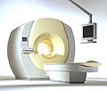
Device Information and Specification CLINICAL APPLICATION Whole body SE, IR, 2D/3D TurboSE, Turbo IR, Dark-Fluid IR, True IR, 2D/3D MEDIC, 2D/3D GRE FLASH, 2D/3D GRE FISP, 2D/3D PSIF, 2D TurboFLASH, 3D MP-RAGE, 3D TurboFLASH, 2D/3D TOF angiography, MTC, TONE with 3D TOF MRA, GMR, LOTA IMAGING MODES Single, multislice, volume study, multi angle, multi oblique178 images/sec at 256 x 256 at 100% FOV1024 x 1024 full screen display POWER REQUIREMENTS 380/400/420/440/480 V Passive, act.; 1st order std./2nd opt. | | | | | |
| | | Searchterm 'mra' was also found in the following services: | | | | |
| | |
| |
|
A type of MRA used to display slow flow across a large volume with a good resolution.
Two data volumes are measured; the flow-rephased images show bright signal, the flow-dephased image show dark flow, whereby in both data volumes the signal of the stationary tissue looks the same. The data volumes are subtracted and the signal intensity of flowing blood remains. | | | | | |
| | | | | |
| |
|
(MIP) MRA images can be processed by Maximum Intensity Projection to interactively create different projections. The MIP connects the high intensity dots of the blood vessels in three dimensions, providing an angiogram that can be viewed from any projection.
Each point in the MIP represents the highest intensity experienced in that location on any partition within the imaging volume.
For complete interpretation the base slices should also be reviewed individually and with multiplanar reconstruction (MPR) software. The MIP can then be
displayed in a CINE format or filmed as multiple images acquired from different projections. Although the maximum intensity projection (MIP) algorithm is sensitive to high
signal from inflowing spins, it is also sensitive to high signal of any other etiology. | | | | | |
• View the DATABASE results for 'Maximum Intensity Projection' (5).
| | | | |  Further Reading: Further Reading: | News & More:
|
|
| |
| | | | |
| | |
| | | |
|
| |
 | Look
Ups |
| |