| Info
Sheets |
| | | | | | | | | | | | | | | | | | | | | | | | |
 | Out-
side |
| | | | |
|
| | | | | |  | Searchterm 'pulse sequences' was also found in the following services: | | | | |
|  |  |
| |
|
Device Information and Specification CLINICAL APPLICATION Whole body Head and body coil standard; all other coils optional; open architecture makes system compatible with a wide selection of coils Standard: SE, IR, 2D/3D GRE and SPGR, Angiography;; 2D/3D TOF, 2D/3D Phase Contrast;; 2D/3D FSE, 2D/3D FGRE and FSPGR, SSFP, FLAIR, optional: EPI, 2D/3D Fiesta, FGRET, SpiralTR 4.4 msec to 12000 msec in increments of 1 msec TE 1.0 to 2000 msec; increments of 1 msec Simultaneous scan and reconstruction;; up to 100 images/second with Reflex 100 2D 0.7 mm to 20 mm; 3D 0.1 mm to 5 mm 128x512 steps 32 phase encode 0.08 mm; 0.02 mm optional POWER REQUIREMENTS 480 or 380/415 V Less than 0.03 L/hr liquid heliumSTRENGTH SmartSpeed 23 mT/m, HiSpeed Plus 33 mT/m 4.0 m x 2.8 m axial x radial | | | | | |
| | | | | |
| |
|
| | | |
• View the DATABASE results for 'Spin Echo' (96).
| | |
• View the NEWS results for 'Spin Echo' (1).
| | | | |  Further Reading: Further Reading: | | Basics:
|
|
News & More:
| |
| |
| | | | | |
| |
|
Magnetic resonance imaging ( MRI) of the spine is a noninvasive procedure to evaluate different types of tissue, including the spinal cord, vertebral disks and spaces between the vertebrae through which the nerves travel, as well as distinguish healthy tissue from diseased tissue.
The cervical, thoracic and lumbar spine MRI should be scanned in individual sections.
The scan protocol parameter like e.g. the field of view ( FOV), slice thickness and matrix are usually different for cervical, thoracic and lumbar spine MRI, but the method
is similar. The standard views in the basic spinal MRI scan to create detailed slices (cross sections) are sagittal T1 weighted and T2 weighted images over the whole body part, and transverse (e.g. multi angle oblique) over the region of interest with different pulse sequences according to the result of the sagittal slices. Additional views or different types of pulse sequences like fat suppression, fluid attenuation inversion recovery ( FLAIR) or
diffusion weighted imaging are created dependent on the indication.
Indications:
•
Neurological deficit, evidence of radiculopathy, cauda equina compression
•
Primary tumors or drop metastases
•
Infection/inflammatory disease, multiple sclerosis
•
Postoperative evaluation of lumbar spine: disk vs. scar
•
Localized back pain with no radiculopathy (leg pain)
Contrast enhanced MRI techniques delineate infections vs. malignancies, show a syrinx cavity and support to differentiate the postoperative conditions. After surgery for disk disease, significant fibrosis can occur in the spine. This scarring can mimic residual disk herniation. Magnetic resonance myelography evaluates spinal stenosis and various intervertebral discs can be imaged with multi angle oblique techniques. Cine series can be used to show true range of motion studies of parts of the spine.
Advanced open MRI devices are developed to perform positional scans in the position of pain or symptom (e.g. Upright™ MRI formerly Stand-Up MRI). | | | | | |
• View the DATABASE results for 'Spine MRI' (11).
| | |
• View the NEWS results for 'Spine MRI' (4).
| | | | | | Further Reading: | | Basics:
|
|
News & More:
| |
| |
| | | Searchterm 'pulse sequences' was also found in the following services: | | | | |
| | |
| |
|
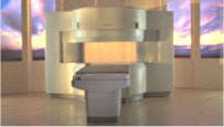
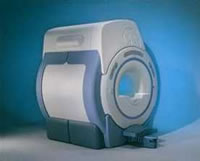
From Hitachi Medical Systems America Inc.;
the AIRIS II, an entry in the diagnostic category of open MR systems, was designed by Hitachi
Medical Systems America Inc. (Twinsburg, OH, USA) and Hitachi Medical Corp. (Tokyo) and is manufactured by the Tokyo branch. A 0.3 T field-strength magnet and phased array coils deliver high image quality without the need for a tunnel-type high-field system, thereby significantly improving patient comfort not only for claustrophobic patients.
Device Information and Specification
CLINICAL APPLICATION
Whole body
QD Head, MA Head and Neck, QD C-Spine, MA or QD Shoulder, MA CTL Spine, QD Knee, Neck, QD TMJ, QD Breast, QD Flex Body (4 sizes), Small and Large Extrem., QD Wrist, MA Foot and Ankle (WIP), PVA (WIP)
SE, GE, GR, IR, FIR, STIR, FSE, ss-FSE, FLAIR, EPI -DWI, SE-EPI, ms - EPI, SSP, MTC, SARGE, RSSG, TRSG, MRCP, Angiography: CE, 2D/3D TOF
IMAGING MODES
Single, multislice, volume study
TR
SE: 30 - 10,000msec GE: 20 - 10,000msec IR: 50 - 16,700msec FSE: 200 - 16,7000msec
TE
SE : 10 - 250msec IR: 10 -250msec GE: 5 - 50 msec FSE: 15 - 2,000
0.05 sec/image (256 x 256)
2D: 2 - 100 mm; 3D: 0.5 - 5 mm
Level Range: -2,000 to +4,000
POWER REQUIREMENTS
208/220/240 V, single phase
COOLING SYSTEM TYPE
Air-cooled
2.0 m lateral, 2.5 m vert./long
| | | |
• View the DATABASE results for 'AIRIS II™' (2).
| | | | |
| | | | | |
| |
|
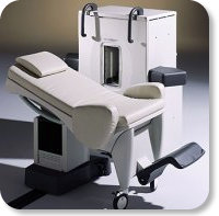
Developed by GE Lunar; the ARTOSCAN™-M is designed specifically for in-office musculoskeletal imaging. ARTOSCAN-M's compact, modular design allows placing within a clinical environment, bringing MRI to the patient. Patients remain outside the magnet at all times during the examinations, enabling constant patient-technologist contact. ARTOSCAN-M requires no special RF room, magnetic shielding, special power supply or air conditioning.
The C-SCAN™ (also known as Artoscan C) is developed from the ARTOSCAN™ - M, with a new computer platform.
Device Information and Specification
CLINICAL APPLICATION
Dedicated extremity
SE, GE, IR, STIR, FSE, 3D CE, GE-STIR, 3D GE, ME, TME, HSE
SLICE THICKNESS
2D: 2 mm - 10 mm;
3D: 0.6 mm - 10 mm
4,096 gray lvls, 256 lvls in 3D
POWER REQUIREMENTS
100/110/200/220/230/240V
| | | |
• View the DATABASE results for 'ARTOSCAN™ - M' (3).
| | | | |
| | | | |
| | | |
|
| |
 | Look
Ups |
| |