 | Info
Sheets |
| | | | | | | | | | | | | | | | | | | | | | | | |
 | Out-
side |
| | | | |
|
| | | | |
Result : Searchterm 'sense' found in 2 terms [ ] and 25 definitions [ ] and 25 definitions [ ] ]
| | previous 6 - 10 (of 27) nextResult Pages : [1] [2 3 4 5 6] |  | |  | Searchterm 'sense' was also found in the following services: | | | | |
| |  |
| |
|
Device Information and Specification
CLINICAL APPLICATION
Whole body
CONFIGURATION
Short bore compact
Standard: head, body, C1, C3; Optional: Small joint, flex-E, flex-R, endocavitary (L and S), dual TMJ, knee, neck, T/L spine, breast; Optional phased array: Spine, pediatric, 3rd party connector, Optional SENSE Coils: Flex-S-M-L, Flex Body, Flex Cardiac
SE, Modified-SE, IR (T1, T2, PD), STIR, FLAIR, SPIR, FFE, T1-FFE, T2-FFE, Balanced FFE, TFE, Balanced TFE, Dynamic, Keyhole, 3D, Multi Chunk 3D, Multi Stack 3D, K Space Shutter, MTC, TSE, Dual IR, DRIVE, EPI, Cine, 2DMSS, DAVE, Mixed Mode; Angiography: Inflow MRA, TONE, PCA, CE MRA
TR
Min. 2.9 (Omni) msec, 1.6 (Power) msec
TE
Min. 1.0 (Omni) msec, 0.7 (Power) msec
RapidView Recon. greater than 500 @ 256 Matrix
0.1 mm(Omni), 0.05 mm (Power)
128 x 128, 256 x 256,512 x 512,1024 x 1024 (64 for Bold img)
Variable in 1% increments
Lum.: 120 cd/m2; contrast: 150:1
Variable (op. param. depend.)
POWER REQUIREMENTS
380/400 V
STRENGTH
23 mT/m (Omni), 30 (Power) mT/m
| | | | | | | | | | |
| | | | | |
| |
|
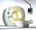
From Philips Medical Systems;
the Intera-family offers with this member a wide range of possibilities, efficiency and a ergonomic and intuitive serving-platform. Also available as Intera CV for cardiac and Intera I/T for interventional MR procedures.
The scanners are also equipped with SENSE technology, which is essential for high-quality contrast enhanced magnetic resonance angiography, interactive cardiac MR and diffusion tensor imaging ( DTI) fiber tracking.
The increased accuracy and clarity of MR scans obtained with this technology allow for faster and more accurate diagnosis of potential problems like patient friendliness and expands the breadth of applications including cardiology, oncology and interventional MR.
Device Information and Specification
CLINICAL APPLICATION
Whole body
CONFIGURATION
Short bore compact
Standard: head, body, C1, C3; Optional: Small joint, flex-E, flex-R, endocavitary (L and S), dual TMJ, knee, neck, T/L spine, breast; Optional phased array: Spine, pediatric, 3rd party connector; Optional SENSE coils: Flex-S-M-L, flex body, flex cardiac
SE, Modified-SE ( TSE), IR (T1, T2, PD), STIR, FLAIR, SPIR, FFE, T1-FFE, T2-FFE, Balanced FFE, TFE, Balanced TFE, Dynamic, Keyhole, 3D, Multi Chunk 3D, Multi Stack 3D, K Space Shutter, MTC, TSE, Dual IR, DRIVE, EPI, Cine, 2DMSS, DAVE, Mixed Mode; Angiography: PCA, MCA, Inflow MRA, CE
TR
2.9 (Omni), 1.6 (Power), 1.6 (Master/Expl) msec
TE
1.0 (Omni), 0.7 (Power), 0.5 (Master/Expl) msec
RapidView Recon. greater than 500 @ 256 Matrix
0.1 mm(Omni), 0.05 mm (Pwr/Mstr/Expl)
128 x 128, 256 x 256,512 x 512,1024 x 1024 (64 for BOLD img.)
Variable in 1% increments
Lum.: 120 cd/m2; contrast: 150:1
Variable (op. param. depend.)
POWER REQUIREMENTS
380/400 V
| | | |
• View the DATABASE results for 'Intera 1.5T™' (2).
| | | | |
| | | | | | | | Searchterm 'sense' was also found in the following services: | | | | |
| | |
| |
|
| | | |
• View the DATABASE results for 'Coronary Angiography' (7).
| | | | |  Further Reading: Further Reading: | | Basics:
|
|
News & More:
| |
| |
| | | | | |
| |
|
(GRAPPA) GRAPPA is a parallel imaging technique to speed up MRI pulse sequences. The Fourier plane of the image is reconstructed from the frequency signals of each coil ( reconstruction in the frequency domain).
Parallel imaging techniques like GRAPPA, auto-SMASH and VD-AUTO-SMASH are second and third generation algorithms using k-space undersampling. A model from a part of the center of k-space is acquired, to find the coefficients of the signals from each coil element, and to reconstruct the missing intermediary lines. The acquisition of these additional lines is a form of self-calibration, which lengthens the overall short scan time. The acquisition of these k-space lines provides mapping of the whole field as well as data for the image contrast.
Algorithms of the GRAPPA type work better than the SENSE type in heterogeneous body parts like thoracic or abdominal imaging, or in pulse sequences like echo planar imaging. This is caused by differences between the sensitivity map and the pulse sequence (e.g. artifacts) or an unreliable sensitivity map. | | | |
• View the DATABASE results for 'Generalized Autocalibrating Partially Parallel Acquisition' (2).
| | | | |
| | | | |
| | |
| | | |
|
| |
 | Look
Ups |
| |


